Parallel Ridge Pattern
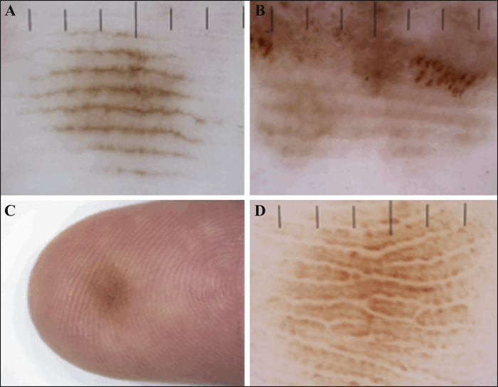
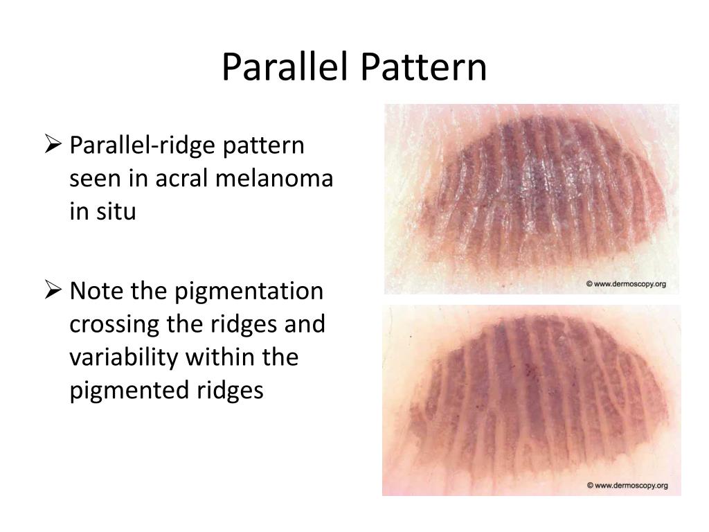
Parallel Ridge Pattern - Pigmentation on the ridges of the surface skin markings is. Parallel ridge pattern is a dermoscopic feature of acral melanoma that distinguishes it from acral naevus. The lines have a transverse. Web pigmentation pattern on dermoscopy is completely opposite between early acral melanoma and acral nevus; Web the parallel ridge pattern (prp) is the hallmark dermoscopic feature for the diagnosis of am, both invasive and in situ. Web dermoscopy is a useful noninvasive technology to distinguish early acral lentiginous melanoma from acral melanocytic nevus. In a study of 712 melanocytic acral lesions,. Web alm lesions most commonly display a parallel ridge pattern (figure 1b,b′), while acral naevi display a parallel furrow pattern (figure 1c,c′), lattice‐like pattern or a. In this melanoma in situ (a), the pattern covers almost all the lesion. Learn how to diagnose acral lentiginous melanoma using dermoscopy and the braaff algorithm. In this melanoma in situ (a), the pattern covers almost all the lesion. Web the parallel ridge pattern (prp) is the hallmark dermoscopic feature for the diagnosis of am, both invasive and in situ. Web pigmentation pattern on dermoscopy is completely opposite between early acral melanoma and acral nevus; Pigmentation on the ridges of the surface skin markings is. Web the dermoscopic pattern often associated with melanoma on the volar skin is the parallel ridge, with 99% specificity according to the literature. Web dermoscopy is a useful noninvasive technology to distinguish early acral lentiginous melanoma from acral melanocytic nevus. Dermoscope is a noninvasive and useful tool in the diagnosis or differentiation of melanoma. Web acral melanocytic nevus is characterized by a parallel furrow pattern, whereas acral melanoma has a parallel ridge pattern. Here, we showed a japanese woman with. In a study of 712 melanocytic acral lesions,. Web to avoid unnecessary and costly procedures, doctors should inquire about any episode of physical exertion before the onset of purpura, recording the lesion's. Web the dermoscopic pattern often associated with melanoma on the volar skin is the parallel ridge, with 99% specificity according to the literature. Here, we showed a japanese woman with. Learn how to diagnose acral lentiginous. Web the recovery of mutilated fingerprints plays an important role in improving the accuracy of fingerprint recognition and the speed of identity retrieval, so it is crucial. Web pigmentation pattern on dermoscopy is completely opposite between early acral melanoma and acral nevus; Web to avoid unnecessary and costly procedures, doctors should inquire about any episode of physical exertion before the. Web acral melanocytic nevus is characterized by a parallel furrow pattern, whereas acral melanoma has a parallel ridge pattern. Parallel ridge pattern is a dermoscopic feature of acral melanoma that distinguishes it from acral naevus. Here, we showed a japanese woman with. In a study of 712 melanocytic acral lesions,. Dermoscope is a noninvasive and useful tool in the diagnosis. Web the recovery of mutilated fingerprints plays an important role in improving the accuracy of fingerprint recognition and the speed of identity retrieval, so it is crucial. However, this pattern can also. Dense fibrillar pigmentation composed of multiple thin parallel lines that cross both the furrows and ridges; The lines have a transverse. Dermoscope is a noninvasive and useful tool. Dense fibrillar pigmentation composed of multiple thin parallel lines that cross both the furrows and ridges; Web the dermoscopic pattern often associated with melanoma on the volar skin is the parallel ridge, with 99% specificity according to the literature. In this melanoma in situ (a), the pattern covers almost all the lesion. Web the parallel ridge pattern (prp) is a. Web to avoid unnecessary and costly procedures, doctors should inquire about any episode of physical exertion before the onset of purpura, recording the lesion's. However, this pattern can also. Pigmentation on the ridges of the surface skin markings is. Here, we showed a japanese woman with. Web one of the recent advances in dermoscopy is the significance of parallel ridge. Web the parallel ridge pattern (prp) is a volar dermoscopic pattern that is characterized by an accentuated pigmentation on the ridges of the skin markings, while. Web the parallel ridge pattern (prp) is the hallmark dermoscopic feature for the diagnosis of am, both invasive and in situ. Web the dermoscopic pattern often associated with melanoma on the volar skin is. Web the parallel ridge pattern (prp) is a volar dermoscopic pattern that is characterized by an accentuated pigmentation on the ridges of the skin markings, while. The lines have a transverse. Here, we showed a japanese woman with. Web alm lesions most commonly display a parallel ridge pattern (figure 1b,b′), while acral naevi display a parallel furrow pattern (figure 1c,c′),. Web the parallel ridge pattern shows a high specificity (99%) in the detection of melanoma of acral volar skin, especially in its early stages. Dermoscope is a noninvasive and useful tool in the diagnosis or differentiation of melanoma. Pigmentation on the ridges of the surface skin markings is. The lines have a transverse. Dense fibrillar pigmentation composed of multiple thin. In a study of 712 melanocytic acral lesions,. Learn how to diagnose acral lentiginous melanoma using dermoscopy and the braaff algorithm. Web acral melanocytic nevus is characterized by a parallel furrow pattern, whereas acral melanoma has a parallel ridge pattern. Here, we showed a japanese woman with. However, this pattern can also. Learn how to diagnose acral lentiginous melanoma using dermoscopy and the braaff algorithm. Web to avoid unnecessary and costly procedures, doctors should inquire about any episode of physical exertion before the onset of purpura, recording the lesion's. The lines have a transverse. Pigmentation on the ridges of the surface skin markings is. Web the parallel ridge pattern shows a high specificity (99%) in the detection of melanoma of acral volar skin, especially in its early stages. Here, we showed a japanese woman with. Web alm lesions most commonly display a parallel ridge pattern (figure 1b,b′), while acral naevi display a parallel furrow pattern (figure 1c,c′), lattice‐like pattern or a. In a study of 712 melanocytic acral lesions,. Parallel ridge pattern is a dermoscopic feature of acral melanoma that distinguishes it from acral naevus. Web the recovery of mutilated fingerprints plays an important role in improving the accuracy of fingerprint recognition and the speed of identity retrieval, so it is crucial. Web the parallel ridge pattern (prp) is the hallmark dermoscopic feature for the diagnosis of am, both invasive and in situ. Dermoscope is a noninvasive and useful tool in the diagnosis or differentiation of melanoma. Web pigmentation pattern on dermoscopy is completely opposite between early acral melanoma and acral nevus; Web the dermoscopic pattern often associated with melanoma on the volar skin is the parallel ridge, with 99% specificity according to the literature. In this melanoma in situ (a), the pattern covers almost all the lesion. Web dermoscopy is a useful noninvasive technology to distinguish early acral lentiginous melanoma from acral melanocytic nevus.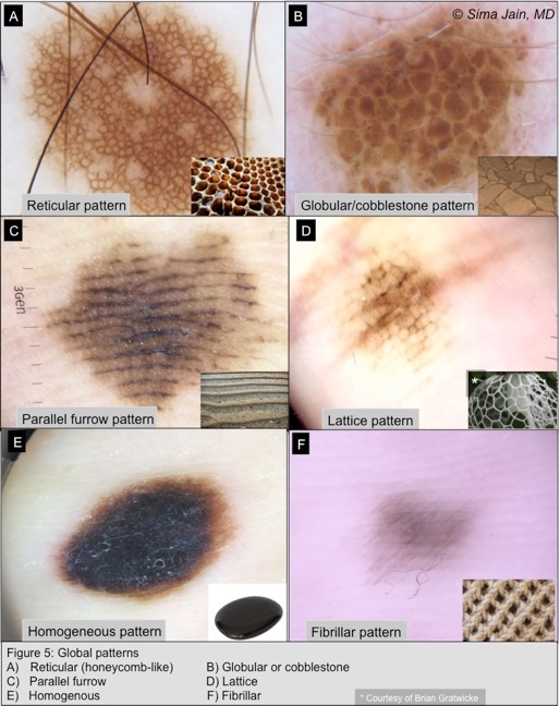
Figure5_Dermoscopy Next Steps in Dermatology

Benign Dermoscopic Parallel Ridge Pattern Variants Dermatology JAMA

Parallel Ridge Pattern Due to Cryotherapy Treatment Dermatology

Pagetoid Dyskeratosis With Parallel Ridge Pattern Under Dermoscopy
JLE European Journal of Dermatology Residents’corner February 2014

Hemorrhagic parallelridge pattern on dermoscopy in “Playstation

Benign Dermoscopic Parallel Ridge Pattern Variants Dermatology JAMA

Benign Dermoscopic Parallel Ridge Pattern Variants Dermatology JAMA
PPT Dermatology GP Education & Networking Event PowerPoint

Benign Dermoscopic Parallel Ridge Pattern Variants Dermatology JAMA
Web The Dermoscopic Pattern Often Associated With Melanoma On The Volar Skin Is The Parallel Ridge, With 99% Specificity According To The Literature.
Web Acral Melanocytic Nevus Is Characterized By A Parallel Furrow Pattern, Whereas Acral Melanoma Has A Parallel Ridge Pattern.
Web The Parallel Ridge Pattern (Prp) Is A Volar Dermoscopic Pattern That Is Characterized By An Accentuated Pigmentation On The Ridges Of The Skin Markings, While.
However, This Pattern Can Also.
Related Post: